Features:
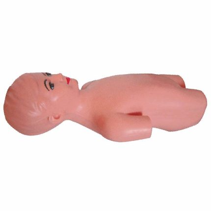
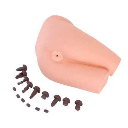
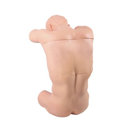
■ Simulation of standardized patients to take the lateral position, back and bed vertical, head bent to the chest, knees bent to the abdomen, the torso is bow-shaped
■ lumbar can move, the operator needs one hand to hold the simulation of the patient’s head, the other hand to hold the two lower limbs of the moon state fossa tight, so that the spine as far as possible to widen the spinal interspace convex, in order to complete the puncture
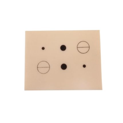
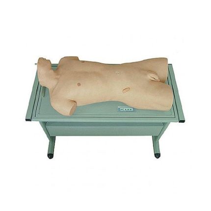
■ Accurate lumbar tissue structure. Body surface markings are obvious: there are complete 1 ~ 5 lumbar vertebrae (vertebral body, arch plate, spinous process), gastric bone, retort fissure, retort angle, supraspinous ligament, interspinous ligament, ligamentum flavum, the dura mater and retina, as well as the formation of the above tissues of the subdural cavity of the retina, the epidural cavity. The retinal canal; the posterior superior spine. The instrumental saki, thoracic spinous processes, lumbar spinous processes can be real palpable
■The following operations are possible: lumbar anesthesia, lumbar puncture, epidural block. Caudal nerve block, retinal nerve block, lumbar sympathetic nerve block
■ lumbar puncture simulation: when the puncture needle arrives at the simulation of the ligamentum flavum, resistance increases with a sense of toughness; break through the ligamentum flavum has a clear sense of emptiness, that is, into the epidural cavity, there is a negative pressure is presented (at this time the injection of anesthetic solution that is the epidural anesthesia); continue to enter the needle will be piercing the dura mater and the retina of the beads, the emergence of the second sense of emptiness, that is, into the subretinal cavity, there will be simulated cerebrospinal fluid outflow, the whole simulation of the clinical lumbar puncture Real plot