Features:
■With all the functions of BOU/F3
■ Hysteroscopy
■ Visual laparoscopy to visualize structures such as the uterus, adnexa, and round ligaments
■ Palpation of the uterus model during pregnancy and after delivery
■ Intrauterine device (IUD) insertion and removal procedure
■ Female condoms can be placed in the vagina, and contraceptive devices such as sponge plugs and cervical caps can be placed and removed.Replaceable cervix and uterus
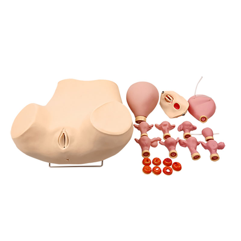
The internal structure of the model consists of the following components:
■ Normal and abnormal uterus and adnexa Models of the normal and abnormal uterus and adnexa, and components for double and triple diagnostic examinations:
-Normal uterus and adnexa (the anterior wall of the uterus is transparent for IUD placement and removal).
-Uterine fibroids
-Uterus with right salpingitis
Uterus with left-sided tubo-ovarian hydrosalpinx
-Uterus with pronounced anterior tilt and flexion.
-Uterus with malformation and right salpingitis.
-Uterus with left ovarian cysts
■ Normal cervix:
-Chronic cervicitis
-Cervical tears
-Cervical polyps
-Inflammatory cervical naboth cysts
-Acute cervicitis
-Cervical adenocarcinoma
■ Normal and abnormal uterine hysteroscopy models (for cervicoscopy components)
-Normal uterus
-Endometrial polyps
-Endometrial hyperplasia
-Uterine fibroids
-Early stage uterine corpus cancer
-Cancer of the uterine corpus, advanced stage
-Cancer of the uterine fundus
■ Pregnancy and postpartum uterine models
-Early pregnant uterus 6-8 weeks
-Early pregnant uterus 10-12 weeks
-Uterus at 20 weeks of pregnancy
-Uterus simulation 48 hours after delivery

